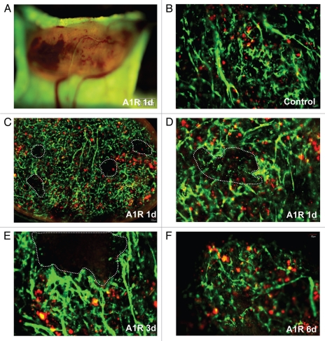
Figure 5.
Bacteria-induced tumor blood vessel destruction in the back. (A) was observed with the Olympus OV100 imaging system. Tumors hemorrhaged near the surface (B–F). Tumors were observed with the Olympus IV100 imaging system. Blood vessels expressed GFP and LLC expressed RFP. Dashed lines show damaged blood vessel areas. In some small areas, tumor blood vessels were damaged at day 1 after A1-R bacteria injection. At days 3 and 6, more blood vessels were damaged.

