Figure 1.
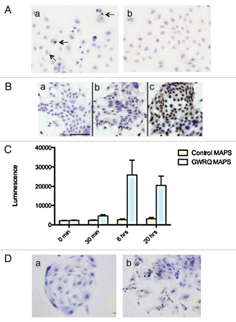
Trophinin-mediated apoptosis of human endometrial epithelial cells. (A) Either trophinin-positive human trophoblastic embryonic carcinoma HT-H cells (a) or control trophinin-negative A431 cells (b) were added to monolayers of human endometrial adenocarcinoma SNG-M cells. Thirty minutes later, cells were removed from the monolayer, and an apoptag TUNEL assay was performed after 24 hours. Arrows in (a) indicate apoptotic nuclei. (B) Human endometrial adenocarcinoma SNG-M cells were subjected to a TUNEL assay 24 hours after adding none (a), control-MAPS (b) or GWRQ-MAPS (c). In all parts, cells were counterstained by hematoxylin. (C) Caspase 3/7 activities were measured using the lysates of SNG-M cells treated with 10 µg/mL each control-MAPS or with GWRQ-MAPS for the indicated times. Each bar and error bar represents mean ± SEM of triplicate measurements. (D) Human endometrial epithelial primary cell cultures were treated with hCG and IL-1β for 6 hours (5) to induce trophinin expression. Cells were subjected to a TUNEL assay 24 hours after addition of control-MAPS (a) or GWRQ-MAPS (b). Scale bars indicate 100 µm.

