Figure 5.
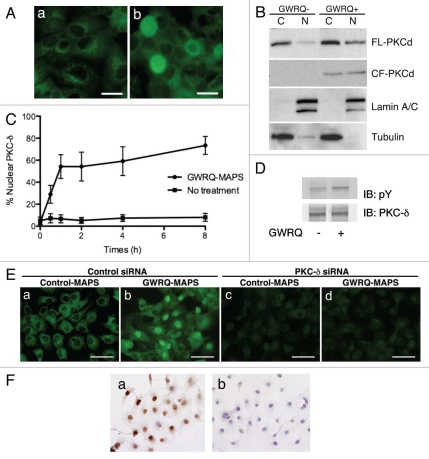
Involvement of PKC-δ in trophinin-mediated apoptosis in human endometrial epithelial SNG-M cells. (A) Immunocytochemistry of PKC-δ in SNG-M cells treated with (b) or without (a) GWRQ-MAPS for 30 minutes. Scale bars indicate 25 µm. (B) Western blot analysis of full-length (FL) PKC-δ and the caspase-3 cleaved form (CF) of PKC-δ distributed to either nuclear (N) or cytoplasmic (C) fractions prepared from SNG-M cells treated with or without GWRQ-MAPS peptide. Lamin and tubulin were included as a control for fractionation. (C) Time-dependency of nuclear translocation of PKC-δ in SNG-M cells cultured in media with or without GWRQ-MAPS. (D) Tyrosine phosphorylation of PKC-δ in SNG-M cells treated with or without GWRQ-MAPS for 30 min. (E) Immunocytochemistry of SNG-M cells by an anti-PKC-δ antibody. SNG-M cells transfected by control (a and b) or PKC-δ (c and d) siRNA were treated with GWRQ-MAPS (b and d) or with control peptide (a and c). Scale bars indicate 50 µm. (F) TUNEL assay of control (a) or PKC-δ (b) siRNA-transfected SNG-M cells treated with GWRQ peptide. A scale bar indicates 50 µm.

