Fig. 3.
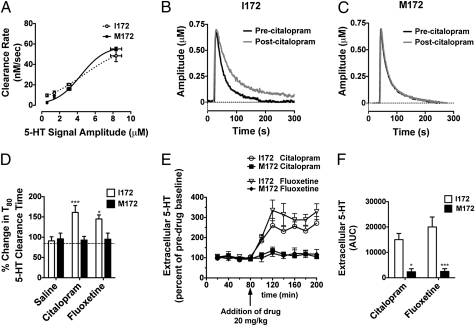
In vivo analysis of SERT-mediated 5-HT clearance and extracellular 5-HT. (A–D) 5-HT clearance measured in the CA3 region of the hippocampus by in vivo amperometry. (A) Baseline clearance rates do not differ between SERT I172 and M172 animals (n = 6 per group; apparent Vmax, 57.7 ± 21.4 nM and 58.6 ± 5.0 nM, respectively). (B) Preapplication of 20 pmol citalopram results in delayed 5-HT clearance in the SERT I172 animals (P < 0.05, Student t test). (C) Preapplication of 20 pmol citalopram has no impact on 5-HT clearance rates in the M172 animals. (D) Percent change in T80 5-HT clearance time (time for signal to decline by 80% of peak signal amplitude) in comparison with baseline T80. I172 SERT mice displayed an increase in the 5-HT T80 clearance time in response to citalopram and fluoxetine, whereas M172 mice did not (*P < 0.05 and ***P < 0.0001 vs. saline solution controls, two-way ANOVA with Bonferroni post-tests; n = 4–8). (E and F) Measurements of extracellular 5-HT by in vivo microdialysis. (E) Time course of the effects of citalopram and fluoxetine administration (20 mg/kg i.p.) on extracellular 5-HT concentrations in the hippocampus of SERT I172 or M172 mice (n = 3–5 for each genotype). SERT I172 mice display increased extracellular levels of 5-HT in response to citalopram, whereas SERT M172 mice do not (P < 0.05, two-way RMANOVA). Values are the mean ± SEM of the percent of predrug baseline. Arrow indicates administration of SSRIs at 80 min. (F) Cumulative elevations of 5-HT across 120-min sample period, expressed as area under the curve, were significantly diminished in the SERT M172 substitution relative to SERT I172 littermate values (*P < 0.05 and ***P < 0.0001 vs. I172, two-way ANOVA with Bonferroni post-tests).

