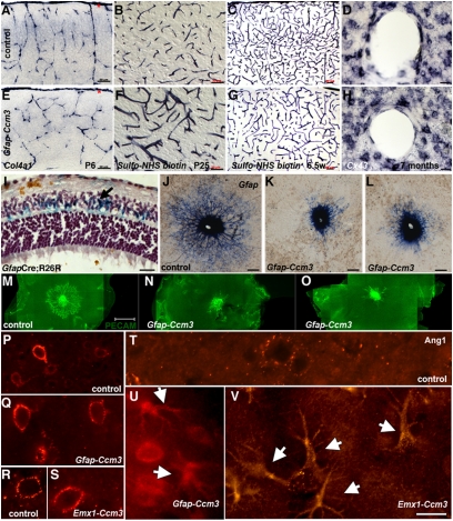
Fig. 3.
Vascular phenotypes in Ccm3 neural mutants. (A and E) Cortical vessels visualized by in situ hybridization for collagen 4a1. Vessels originating from the pial surface (red asterisks) and invading the cortex seem disorganized in Gfap-Ccm3 mutants (E) compared with controls (A). Results are representative of three paired observations. (B, C, F, and G) Cerebral vasculature visualized after intracardiac administration of N-hydroxysulfosuccinimide (sulfo-NHS)-biotin. In Gfap-Ccm3 mutants (F and G), the vessels are dilated (F), and the vascular tree is overall less complex (G) compared with controls (B and C). Results are representative of seven paired observations. (D and H) In situ hybridization staining for connexin 43 (Cx43) indicates lower levels of Cx43 mRNA expression in perivascular astrocytes of Gfap-Ccm3 mutants (H) compared with controls (D). Results are representative of four paired observations. (I) X-gal histochemistry of retina from a 3-wk-old Gfap-Cre animal carrying the R26R reporter (GfapCre;R26R) shows that retinal astrocytes (blue and arrow) derive from Gfap-expressing progenitors. (J–L) In situ hybridization for Gfap in P2 retinas from one control (J) and two Gfap-Ccm3 (K and L) animals. In control retinas (J), astrocytes emerging from the optic nerve spread in a centrifugal fashion; in mutants (K and L), astrocyte spreading is attenuated. Results are representative of six paired observations. (M–O) Immunostaining for platelet endothelial cell adhesion molecule 1 (PECAM-1) in P1 retinas from one control (M) and two Gfap-Ccm3 mutants (N and O) shows retinal endothelial cells following the astrocytic network in controls (M) but are stalled in Gfap-Ccm3 retinas (N and O). Results are representative of four paired observations. (P–V) Immunostaining for Angiopoietin-1 (Ang1) shows expression around cortical vessels and neurons in 7.5-wk-old control (P) and Gfap-Ccm3 (Q) littermates and in 12-mo-old control (R) and Emx1-Ccm3 (S) littermates. Expression in cortical astrocytes is low to undetectable in 12-mo-old controls (T) but dramatically up-regulated in 5.5-mo-old Gfap-Ccm3 (U) and 12-mo-old Emx1-Ccm3 mutants (arrows indicate activated astrocytes of characteristic morphology). Results are representative of three paired observations per genotype at different ages. (Scale bars: A–D, G, and H, 50 μm; E, F, and I, 20 μm; J–L, 200 μm; M–O, 500 μm; P–V, 20 μm.)

