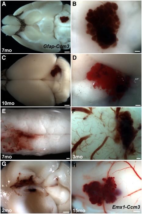
Fig. 4.
Vascular lesions develop in Ccm3 neural mutants. (A–E) Lesions resembling human cavernomas develop in the brains (A–D) and spinal cords (E) of Gfap-Ccm3 animals. B and D are higher magnification views of A and C. (F) Lesion in a 3-mo-old Gfap-Ccm3 animal that died accidentally. (G) Lesion in the dorsomedial cortex of a 2-mo-old animal with severe hydrocephalus, indicated by the collapsed cortical wall. (H) Cortical lesion in a 15-mo-old Emx1-Ccm3 animal. Brains in F and H were extracted fresh without perfusion; cerebral surface vessels can be seen feeding into the lesion. (Scale bars: A and C, 1 mm; B, D, E, and F, 200 μm; G and H, 500 μm.)

