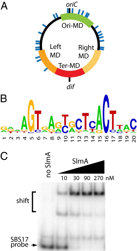
Fig. 2.
Identification of chromosomal SlmA-binding sites. (A) Circular diagram of the E. coli chromosome with approximate locations of SlmA-binding sites shown as blue lines. Green, red, dark- and light-orange colored regions correspond to the Ori, Ter, Left, and Right macrodomains (MDs), respectively (26). (B) Sequence logo of consensus SlmA-binding sequence generated using weblogo. (C) The indicated concentrations of SlmA were incubated with unlabeled poly-dIdC (100 μg/mL) and 0.2 nM [32P]-labeled SBS17-containing DNA (100 bp). The SBS17 site was centered within the probe fragment and flanked by adjacent chromosomal sequence. Protein–DNA complexes were separated from free probe by gel electrophoresis and detected using a phosphorimager. We do not currently know why an intermediate shifted complex is observed.

