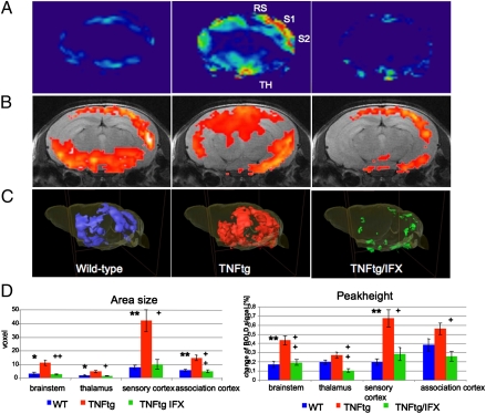
Fig. 3.
Reversible enhancement of central pain responses by TNF-α–mediated arthritis. Functional MRI of the brain of 10-wk-old WT and human TNFtg mice without and with human antiTNF-α antibody IFX (each n = 10). (A and B) 2D axial scans with heatmap (A) or superimposed on the corresponding anatomical image (B) show enhanced neuronal activity in the primary (S1) and secondary (S2) somatosensoric cortex, as well as in association cortex (RS) and the thalamus (Th) of TNFtg mice, which is reversible 24 h after IFX administration. (C) 3D reconstruction of functional brain activity. (D) Bar graphs showing area size (Left) and peak height (Right) of the BOLD signal in WT mice (blue) and TNFtg mice treated with either vehicle (red) or the human anti–TNF-α antibody IFX (green). Four key groups of brain regions (brainstem, thalamus, sensory cortex, and association cortex) involved in central nociception are shown. Asterisks indicate significant differences of WT to TNFtg plus those of TNFtg/IFX to TNFtg, with * and + indicating a P value of < 0.05 and ** and ++ a P value of < 0.025) (n = 10).

