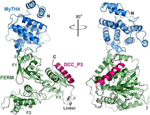
Fig. 2.
Ribbon representation of the MyoX_MF/DCC_P3 complex structure. The color coding of the domains is used throughout the entire manuscript except as otherwise indicated. The linker region contains the residues of the N-terminal part of DCC_P3 (residues 1409–1421). A disordered loop in the F3 lobe and a truncated region (marked by a red arrow) in the F2 lobe are indicated by dashed lines.

