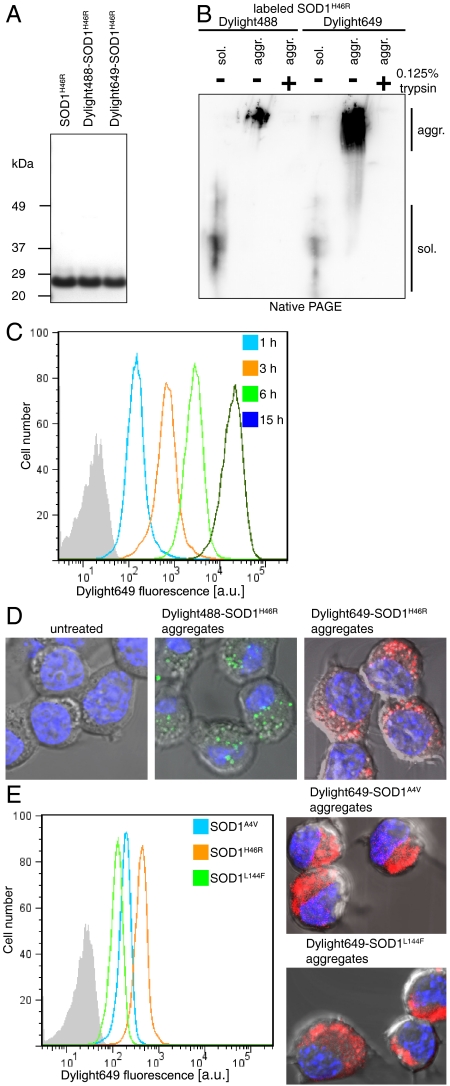
Fig. 1.
Mutant SOD1 aggregates penetrate inside neuronal cells. (A) Purified SOD1H46R (1.2 μg), unlabeled or labeled with Dylight dyes, analyzed by NuPAGE and stained with Coomassie brilliant blue. (B) Soluble (sol.) or aggregated (aggr.) SOD1H46R labeled with Dylight dyes, digested with 0.125% trypsin for 1 min where indicated, prior to native PAGE followed by immunoblot analysis with SOD1 polyclonal antibodies. (C) Neuro-2a take up labeled SOD1H46R aggregates in a time-dependent manner. Flow cytometry analysis of Neuro-2a cells inoculated with Dylight649-SOD1H46R aggregates (0.2 μM monomer equivalent); a.u., arbitrary units. (D) Confocal micrographs of cells untreated or inoculated with labeled SOD1H46R aggregates 15 min before fixing the cells. Nuclei were stained with H33258. Aggregates were estimated to be 2–4 μm inside the cell, not at the surface. Representative results of at least three independent experiments are shown. (E) Flow cytometry and confocal analysis of cells left untreated for 3 h after inoculation with Dylight649-labeled SOD1A4V, SOD1H46R, or SOD1L144F aggregates.

