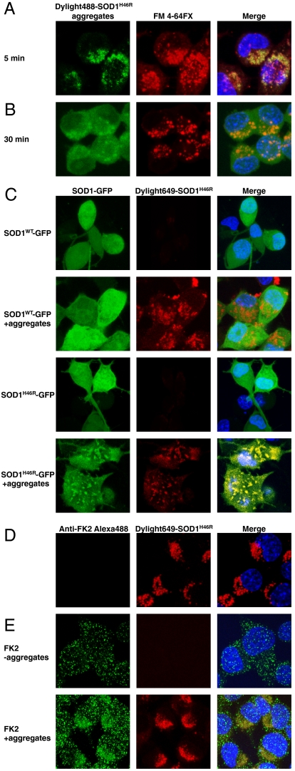
Fig. 2.
Mutant SOD1 aggregates are internalized by vesicles and escape the endocytic compartment to seed aggregation of the endogenous mutant protein. (A) SOD1H46R aggregates localize in membrane-enclosed vesicles. Confocal micrographs of cells after inoculation with Dylight488-SOD1H46R aggregates (green) for 5 min together with the fixable membrane dye FM 4-64FX (red). Nuclei were stained with H33258 dye. (B) Aggregates rapidly exit the FM 4-64FX marked vesicles. Same as in A except that cells were fixed 30 min after addition of aggregates and the FM 4-64FX dye. (C) Confocal micrographs of cells transiently expressing SOD1-GFP wild-type or SOD1H46R-GFP and inoculated with 0.2 μM (monomer equivalent) Dylight649-SOD1H46R aggregates, where indicated, for 15 h before fixing the cells. (D and E) Cells were exposed to Dylight649-SOD1H46R for 15 h, trypsinized, and cultured for 2 d before fixing, labeling with the polyubiquitin antibody FK2 where indicated, and confocal microscopy. Representative results of at least three independent experiments are shown.

