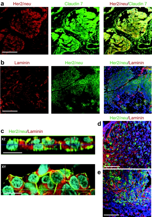
Figure 1.
Immunohistochemical colocalization of Her2/neu and ECM proteins in breast cancer. (a) Representative sections of a tumor biopsy from a patient with stage III ductal mammary carcinoma. (b) Sections of a biopsy from a patient with stage IV clear cell ovarian cancer. Bar = 40 µm. (c) Confocal microscopy of BT474-M1 tumor cells in vitro. Shown are representative images of stacked XZ and XY sections. Bar = 20 µm. (d,e) Sections of xenograft tumors derived from (d) BT474-M1 and (e) HCC1954 cells. Bar = 40 µm. ECM, extracellular matrix.

