Figure 4.
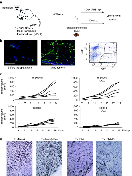
Studies with transplanted mouse HSCs in neu-tg mice carrying MMC tumors. (a) Scheme of experiment. A total of 5 × 10⁵ of mock- or LV-transduced HSCs were transplanted into lethally irradiated neu-tg mice via tail vein injection. Six weeks after HSCs engraftment, MMC tumors were established via injection of 5 × 10⁵ MMC cells subcutaneously. Mice received intraperitoneal injection of PBS or Dox (0.5 mg/mouse in 500 µl PBS) starting at day 7 after MMC cell transplantation and then every other day. (b) Tumor homing of gene modified cells. Left panel: GFP expression in HSCs before transplantation, middle panel: representative MMC tumor section from mice that received LV-GFP transduced HSCs. Right panel: F4/80 and GFP flow cytometry analyses of MMC tumors from mice that received LV-GFP transduced HSCs. Tumors were digested with collagenase to generate single cell suspensions. The gated sections P4 and P5 represent GFP+/F4/80+ and GFP+/F4/40− cells, respectively. Shown is a representative sample. (c) Therapy study with mice that received mock-transduced (upper panels) and Ins-SIN-LV-Rlx-transduced (lower panels) mouse HSCs. Dox or PBS was injected intraperitoneally at day 7 after MMC cell implantation and then every other day. Rlx/Dox− versus Rlx/Dox+: P = 0.0021 for day 19. The P value has been calculated based data from three independent experiments with different numbers of inoculated tumor cells in each experiment. The figure shows the data from animals injected with 5 × 10⁵ MMC cells. (d) Representative sections stained for basement membrane using Jones' periodic acid silver staining method. Basement membrane appears in dark brown/black. Dox, doxycycline; GFP, green fluorescent protein; HSC, hematopoietic stem cell; LV, lentivirus; MMC, mammary carcinoma cell; PBS, phosphate-buffered saline; Rlx, relaxin.

