Figure 7.
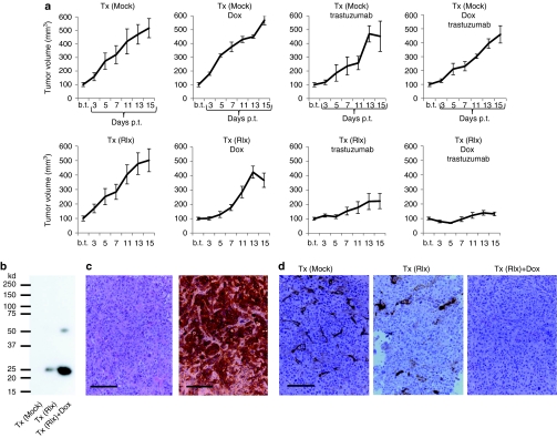
Therapy studies in the HCC1954 model. Mice were treated as described in Figure 6. (a) Tumor growth in mice. Shown is the relative increase of tumor volume. Tumor volumes at the day Dox/PBS injection were started (“b.t.”) were taken as 100%. b.t., before treatment; p.t., post-treatment. N = 5. Shown are the average tumor volumes and SD. In the Tx(Rlx)Dox group, the decrease in tumor volume between days 13 and 15 is not significant. (b) Tumor lysates were subjected to immunoprecipitation with polyclonal anti-Rlx antibodies and analyzed by western blot with monoclonal anti-Rlx antibodies. Representative samples are shown. In agreement with the manufacturer, the monoclonal anti-Rlx antibody reacts with 25 kd and ~50 kd Rlx forms. (c) Representative paraffin sections of tumors from mice without Rlx expression [Tx (Mock)] stained with H&E (left panel) and for Her2/neu (right panel). Tumor sections for the other experimental groups were similar. (d) Immunohistochemistry for collagen IV. Positive signals appear in brown. Bar = 40 µm. Dox, doxycycline; GFP, green fluorescent protein; H&E, hematoxylin and eosin; HSC, hematopoietic stem cell; LV, lentivirus; PBS, phosphate-buffered saline; Rlx, relaxin; SIN, self-inactivating.

