Figure 2.
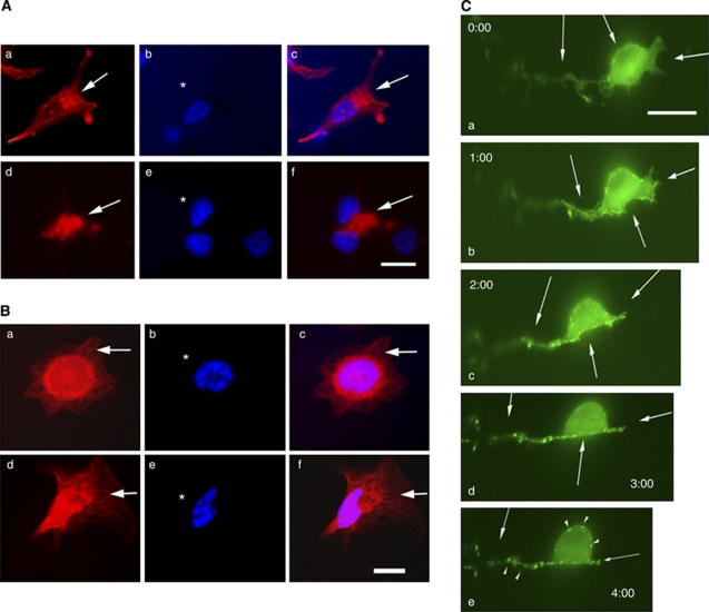
Cuc IIa alters actin cytoskeleton but not microtubule organisation. (A) Cuc IIa induces a dramatic actin clustering in cultured CWR22Rv-1 cells. (a–c) Cells without Cuc IIa treatment. (d–f) Cells treated with 50 μg ml−1 Cuc IIa. Arrows indicate rhodamine phalloidin staining of F-actin. Bar: 20 μm. (B) Cuc IIa does not induce changes in microtubule organisation. (a–c) Cells without Cuc IIa treatment. (d–f) Cells treated with 50 μg ml−1 Cuc IIa. (C) Time-lapse imaging of NIH3T3 cells transfected with EGFP-actin shows gradual aggregation of actin cytoskeleton and the alteration is not reversible. The cells were grown to 60% confluency and were then transfected with EGFP-actin. The cells were treated with 50 μg ml−1 Cuc IIa over a 2-h period followed by a 2-h recovery. Time-lapse images were taken every 5 min. After 2 h of Cuc IIa treatment, the drug was removed and cells were rinsed. The cells were then recorded for an additional 2 h to assess the recovery of actin distribution. Arrows indicate points of increasing actin aggregation with time. Bar: 15 μm.

