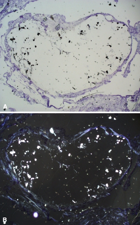
Fig. 5A–B.
Histologic analysis of the synovial lining of a hip spacer from a 71-year-old man shows a greater piece of cement (1.2 mm) with smooth, rounded surfaces as a sign of abrasion (Stain, toluidine blue; original magnification, ×50). (A) With light microscopy, zirconium particles of the cement are dark and (B) can be seen better as light particles with polarized light microscopy.

