
Figure. 1.
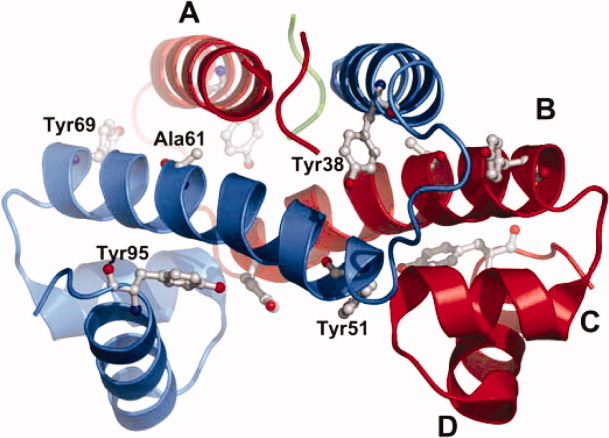
Ribbon model representation of the X-ray crystal structure of P61A FIS (PDB file: 1FIP). The first 25 residues of each monomer are flexible and do not resolve in the crystal structure. The tyrosine residues are highlighted and the four α-helices are labeled A through D. This figure was prepared using the software PyMol (The PyMol Molecular Graphics System, Version 1.3, Schrodinger, LLC.).