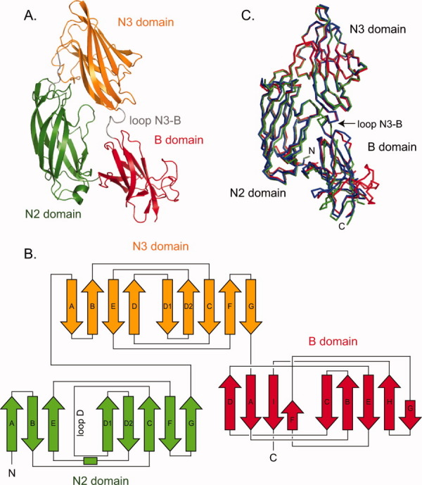
Figure. 3.

Crystal structure of functional region of UafA. (A) Overall structure of UafA-F(392-811). N2, N3, and B domains are shown in green, orange, and red, respectively. The N3-B loop is shown as a gray loop. (B) Topology diagrams of UafA(392-811). Arrows and boxes represent β-strands and α-helixes, respectively. The colors correspond to those in Figure 3(A). (C) Superposition of three structures. UafA-F(376-811), P21 form UafA-F(392-811) (1.5 Å resolution), and P212121 form UafA-F(392-811) (1.7 Å resolution) are shown in blue, red, and green, respectively. Cα atoms in N3 domains are superposed.
