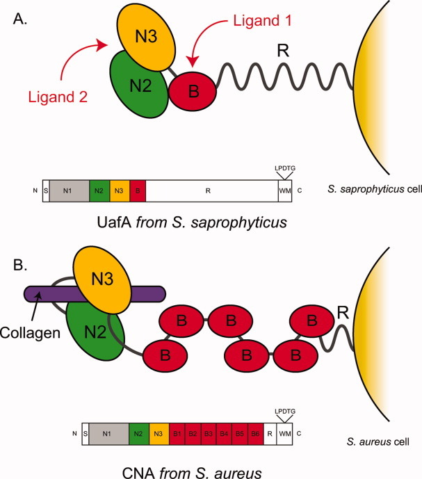
Figure. 7.

Hypothetical ligand binding model of UafA. The ligand binding mechanism of UafA (A, present study) and that of CNA (B16) are shown. N2, N3, and B domains are shown in green, yellow, and red, respectively. Collagen, which is a ligand of CNA, is also shown as a purple bar. The domain organization of each protein is also shown.
