
Figure. 1.
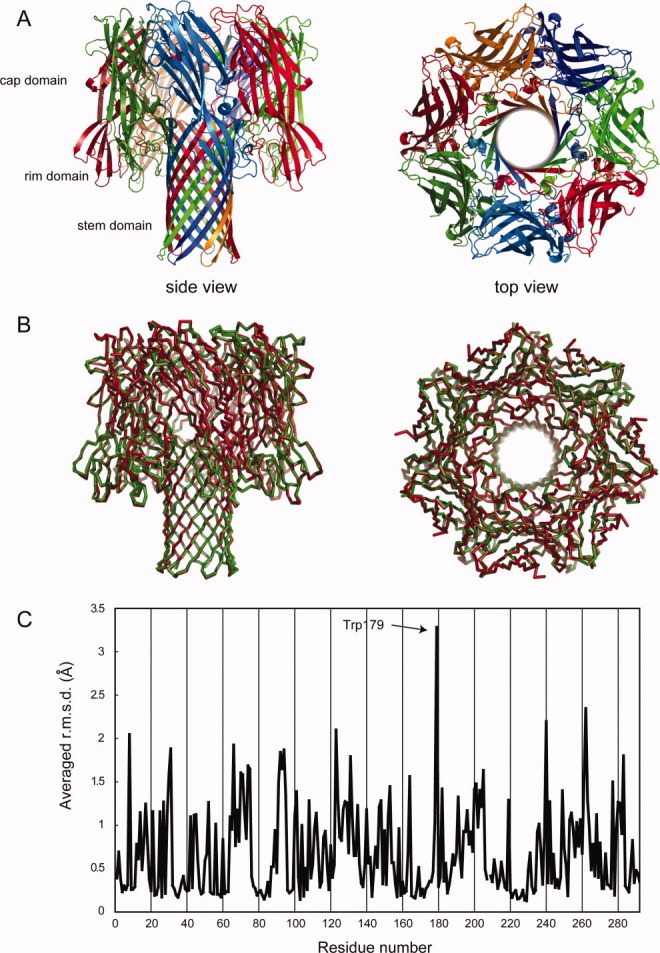
The revealed heptameric structure of α-hemolysin. (A) Overall structure of heptameric structure of α-hemolysin from side (left) and top (right). Protomers are colored individually. (B) Superposition of the heptamer determined in this study (red) and that prepared by deoxycholate (green). Cα traces are shown. (C) The averaged r.m.s.d. of all atoms in each residue between heptamers shown in Fig. 1B. A heptamer was superposed onto the heptamer reported by Song et al. (PDB ID: 7AHL). The r.m.s.d. values are displayed as per-residue averages.