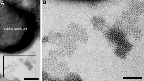
Figure 3.
Electron micrograph of a negatively stained preparation of B. subtilis 1012 cells carrying plasmid pHT43/sbpA/bet v1, 3 h after induction of rsbpA/bet v1 expression. (A) After secretion into the culture medium, the heterologously produced S-layer/allergen fusion protein did not recrystallize on the cell surface of B. subtilis 1012 but was able to form self-assembly products with a maximum size of 1 μm which clearly exhibited the square S-layer lattice symmetry. Bar = 100 nm (B) Detailed view on the ultrastructure of the rSbpA/Bet v1 self-assembly products which show a center-to-center spacing of the morphological unit of 13.1 nm. Bar = 50 nm.

