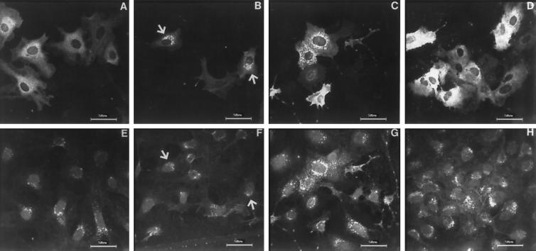
Figure 3.
Immunostaining of OK cells transiently transfected with hmunc13-HA (A–C and E–G) and C1-less mutant (D and H). Cells were stained with anti-HA, then probed with anti-mouse IgG–rhodamine for detection of hmunc13 (A–C) and C1-less mutant (D). The Golgi apparatus was detected by staining with WGA–FITC (E–H). Slides were observed by confocal microscopy using a laser scanning microscope with excitation wavelength at 568 nm for detecting rhodamine (A–D) and at 488 nm for detecting FITC (E–H). Cells were treated with vehicle (A and E), 0.1 μM PDBu for 3 h (B, D, F and H), 4 μM nocodazole + PDBu (C and G) as described in MATERIALS AND METHODS. Negative controls obtained by incubating with normal mouse IgG or immunostaining of cells transfected with empty plasmid (pCMV·SPORT) yielded very little or no staining (our unpublished results). Arrowheads indicate colocalization of anti-HA and WGA staining. Note: top and bottom panel pairs, i.e., A and E, B and F, etc., represent anti-HA and WGA-FITC staining, respectively, of identical fields.

