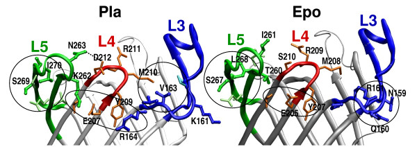
Figure 5.
Analysis of the ten substituted residues in Pla structure (left) and Epo model (right). L3 and its residues are colored blue, L4 red, and L5 green. The substituted residues are drawn thick and in a darker shade, and the other discussed residues thin and in lighter shade. The hydrogen bond between D212 and N263 in Pla is marked with a dashed line, and three interaction areas circled in both structures. Counting from left: a tight cluster of hydrophobic residues in L5; a polar quintet between L5, L4 and L3 in Pla and a L4 triplet in Epo; hydrophobic contacts within the barrel opening in Pla and polar residues at the outside of the barrel in Epo.

