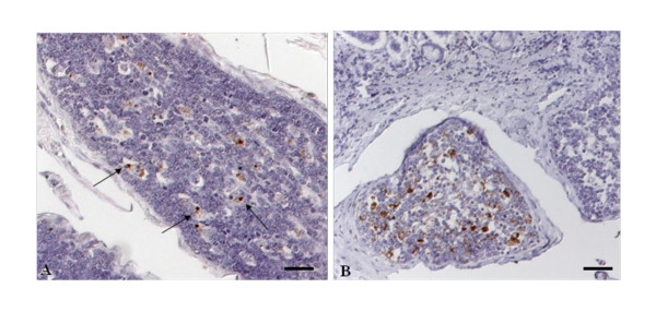
Figure 3.
PrPSc accumulation within follicles of the ileal PP. A: IT55 (8 months p.i.), note the mild, predominantly punctate (arrows) reaction pattern within the cytoplasm of tingible body macrophages only; B: IT24 (24 months p.i.), besides a intracytoplasmatic globular reaction pattern within the TBM's, a clear net-like staining reaction typical for FDC can be seen; Immunohistochemistry, PrP mAb 12F10, Nomarski interference contrast, Bars 50 μm.

