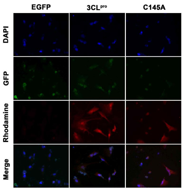
Figure 3.
Nuclear and cytoplasmic localization of 3CLpro and C145A in transfected A549 cells. EGFP, 3CLpro, and C145A transfected A549 cells were collected at 24 hrs after transfection. Cells were fixed and stained as described in Methods. Nuclei were stained with DAPI (blue fluorescence, obtained from the excitation of 359 nm and emission of 468 nm.). GFP panel shows the green fluorescence obtained from the excitation of 488 nm and emission of 509 nm. 3CLpro and C145A were detected with a rhodamine-conjugated secondary antibody (red fluorescence, obtained from the excitation of 496 nm and emission of 520 nm).

