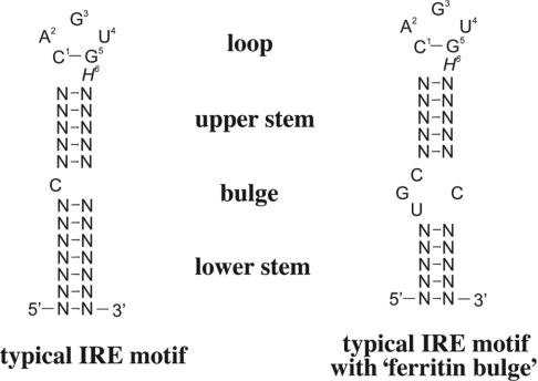
Figure 3. Typical IRE motif.
A typical IRE motif consists of a hexanucleotide loop with the sequence 5′-CAGUGH-3′ (H could be A, C, or U) and a stem, interrupted by a bulge with an unpaired C residue (left) or an asymmetric tetranucleotide bulge (right); the latter is characteristic of ferritin IRE. Base-pairing between C1 and G5 of the loop is functionally important.

