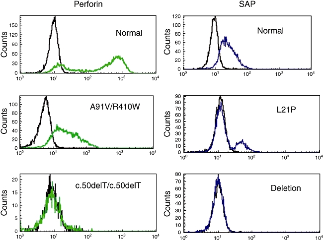
Fig. 2.

Fluorescence activated cell sorter (FACS) base diagnosis of X-linked lymphoproliferative disease (XLP) and familial haemophagocytic lymphohistiocytosis (FHL)2. FACS plots on the left are gated on natural killer (NK) cells and show a normal individual on top and two individuals with confirmed perforin mutations below. FACS plots of the right gated on CD8+ T cells show a normal individual on top and two confirmed XLP patients below. Mutations are indicated in each plot.
