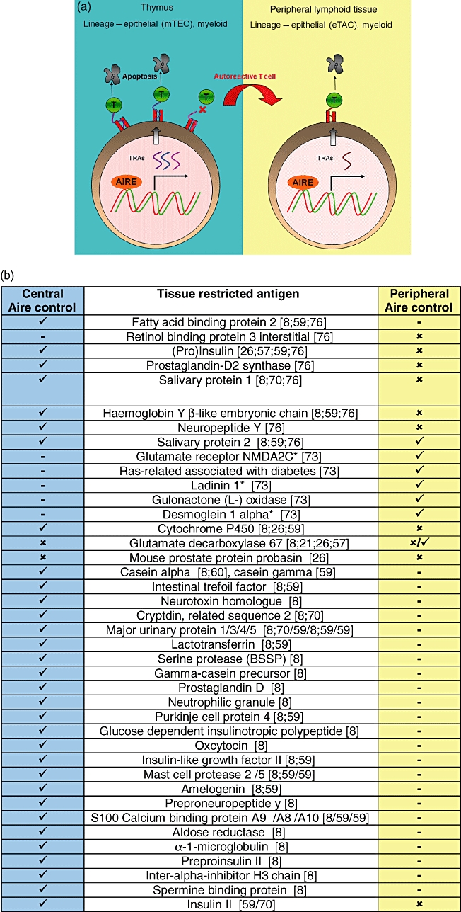
Fig. 2.

Autoimmune regulatory (Aire) protein and TRAs in the thymus and periphery. (a) Cartoon of Aire expression and function in the thymus and peripheral lymphoid tissue. TRAs are expressed by medullary thymic epithelial cells (mTECs) and myeloid cells. T cells which recognize these TRAs with too high affinity/avidity die by apoptosis. Self-reactive T cells which do not interact with their cognate antigen escape to the periphery and are eliminated following exposure to TRAs displayed in the periphery. Aire expressed in the periphery may control the expression of different TRAs to those under Aire control in the thymus. TRAs: tissue restricted antigens; DCs: dendritic cells; ✓: expressed; ✗: not expressed;  : not done. (b) Comparison of thymic and peripheral Aire-restricted TRAs. *Human homologues of these mouse proteins have been described as autoantigens in human autoimmune diseases.
