
Fig. 1.
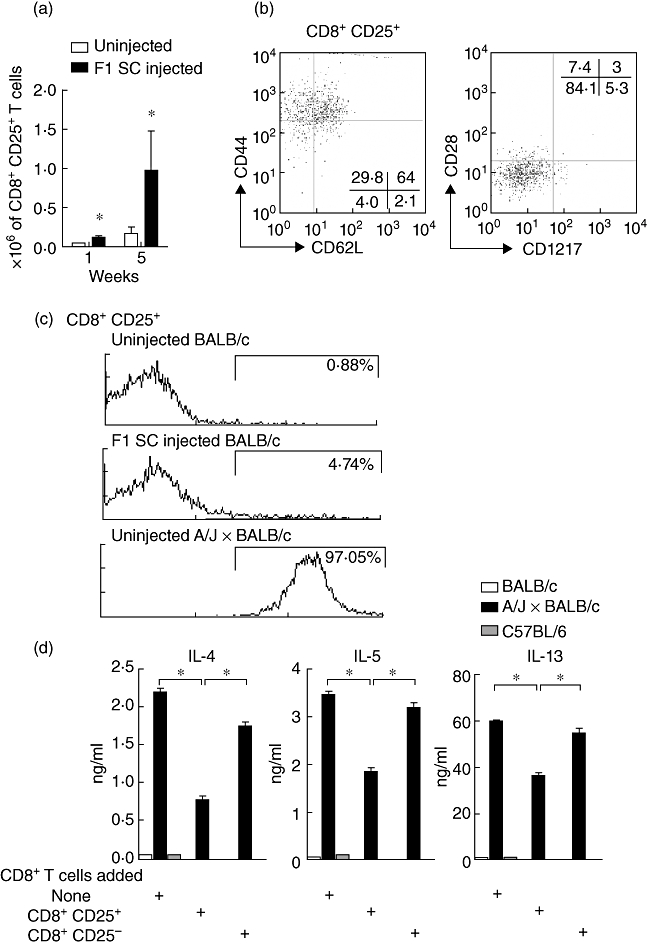
Phenotype of CD8+ T cells in F1 spleen cells neonatally injected mice and inhibition of T helper type 2 (Th2) cytokine production. (a) Absolute numbers of CD8+CD25+ T cells in total lymph nodes collected from uninjected and neonatally injected mice at 1 and 5 weeks of age. Results are mean values from five separate experiments, each involving a pool of five mice (*P < 0·004 compared with uninjected mice at 1 week of age and *P < 0·05 compared with uninjected mice at 5 weeks of age). (b) CD44/CD62L staining or CD28/CD127 staining on gated CD8+CD25+ T cells. Results are expressed as % of positively stained cells and are representative of three separate experiments, each involving a pool of five mice. (c) H-2Kk expression on CD8+CD25+ T cells collected from 5-week-old uninjected BALB/c mice, F1 spleen cells injected mice or donor uninjected A/J × BALB/c mice (pool of seven mice). (d) In vitro suppressive activity of purified CD8+CD25+ T cells versus purified CD8+CD25- T cells on Th2 cytokine production by CD4+ T cells from neonatally injected BALB/c wild-type mice; 2·5 × 106 CD8+ T cell subsets were cultured with 2·5 × 106 Th2 cytokine-producing CD4+ T cells and 2 × 10⁶ irradiated allogeneic A/J × BALB/c spleen cells for 72 h or when indicated with 2 × 10⁶ irradiated syngeneic BALB/c or third-party C57BL/6 spleen cells. Results are expressed as ng/ml of interleukin (IL)-4, IL-5 and IL-13 measured by enzyme-linked immunosorbent assay (ELISA). Results are representative of three individual experiments containing five mice per group (*P < 0·05).