
Fig. 3.
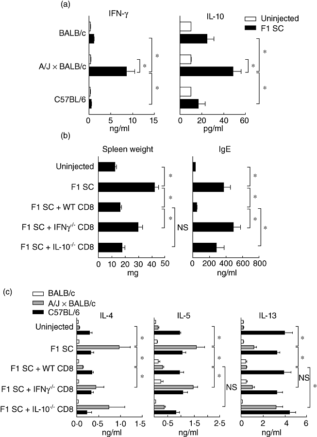
Role of interferon (IFN)-γ and interleukin (IL)-10 produced by CD8+ T cells in the control of the T helper type 2 (Th2) pathology. (a) Purified CD8+ T cells from uninjected and neonatally injected mice were cultured with irradiated syngeneic (BALB/c), allogeneic (A/J × BALB/c) or third-party (C57BL/6) spleen cells. Supernatants were collected after 72 h of culture and the presence of IFN-γ and IL-10 was determined by enzyme-linked immunosorbent assay (ELISA). Results are mean values of six pools of three mice per experimental group (*P < 0·05). (b,c) β2m−/− BALB/c mice were injected at birth with (A/J × BALB/c) F1 spleen cells and wild-type, IFN-γ−/− or IL-10−/− CD8+ T cells purified from neonatally injected BALB/c mice. (b) Spleen weight expressed in mg and seric immunoglobulin (Ig)E expressed in ng/ml monitored at 4 weeks of age. Results are mean values of at least 10 mice per group (*P < 0·05). (c) Total lymph node (LN) cells were cultivated with irradiated syngeneic (BALB/c), allogeneic (A/J × BALB/c) or third-party (C57BL/6) spleen cells. Supernatants were collected after 72 h and the presence of IL-4, IL-5 and IL-13 were determined by ELISA. Results are mean values of five to 14 pools of three mice per experimental group (*P < 0·05); n.s.: non-significant.