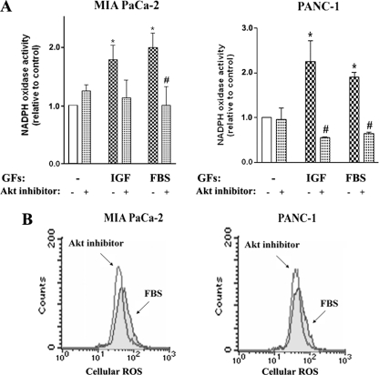
FIGURE 4.
Akt inhibition prevents stimulation of NADPH oxidase by growth factors. MIA PaCa-2 and Panc-1 cells were cultured for 48 h in the absence or presence of IGF-I (100 ng/ml), FBS (15%), or the Akti-1/2 inhibitor (50 μm). A, superoxide production was measured by lucigenin-derived chemiluminescence in total cell homogenate. The chemiluminescence values were normalized to those in cells cultured in the absence of the GFs and Akt inhibitor. Values are means ± S.E. (n = 3). *, p < 0.05 versus cells cultured in the absence of the GFs and Akt inhibitor. #, p < 0.05 versus cells cultured in the same conditions but without Akt inhibitor. B, changes in intracellular ROS level were measured by FACS® analysis using the redox-sensitive dye DCFH-DA in cells cultured with FBS. Data are representative of two independent experiments that gave similar results.

