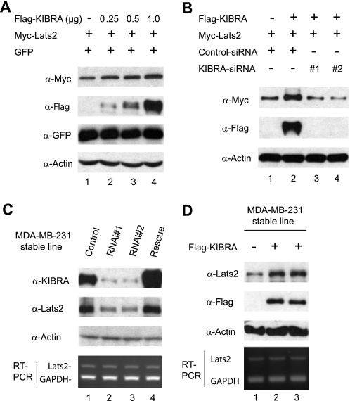
FIGURE 5.
Effects of KIBRA expression on Lats2 protein levels. A, HEK293T cells were transfected with Myc-tagged Lats2 and pEGFP-C1(GFP) with or without increasing amounts of FLAG-tagged KIBRA. At 48 h after transfection, total cell lysates were subjected to Western blot analysis to detect the Lats2 and GFP protein levels. The lysates were also probed with anti-FLAG to detect the KIBRA expression, and with anti-β-actin antibody to show equal loading. B, HEK293T cells were transfected with Myc-Lats2 with (lanes 2–4) or without (lane 1) FLAG-KIBRA together either with siRNA oligonucleotides targeting the human KIBRA sequence (lanes 3 and 4) or with a scrambled oligonucleotide (lanes 1 and 2). At 48 h post-transfection, total cell lysates were subjected to Western blot analysis using indicated antibodies. C, MDA-MB-231 cell lines stably expressing control shRNA or shRNA targeting human KIBRA were established (see “Experimental Procedures”). The rescue cells were generated as in Fig. 3. Total protein lysates were subjected to Western blot analysis with anti-Lats2 and anti-KIBRA antibodies to detect the Lats2 and KIBRA protein levels, respectively. Two pooled lines are shown. Semiquantitative RT-PCR was performed with primers amplifying Lats2 and GAPDH (control) (bottom panel). D, MDA-MB-231 cell lines stably expressing control vector or FLAG-KIBRA were established (see “Experimental Procedures”). Total protein lysates were subjected to Western blot analysis with anti-Lats2 antibody to detect the endogenous Lats2 protein level and with anti-FLAG antibody to confirm KIBRA expression (two separately generated lines are shown). Total RNA was also isolated from these cell lines and semiquantitative RT-PCR was performed with primers amplifying Lats2 and GAPDH (control) (bottom panel).

