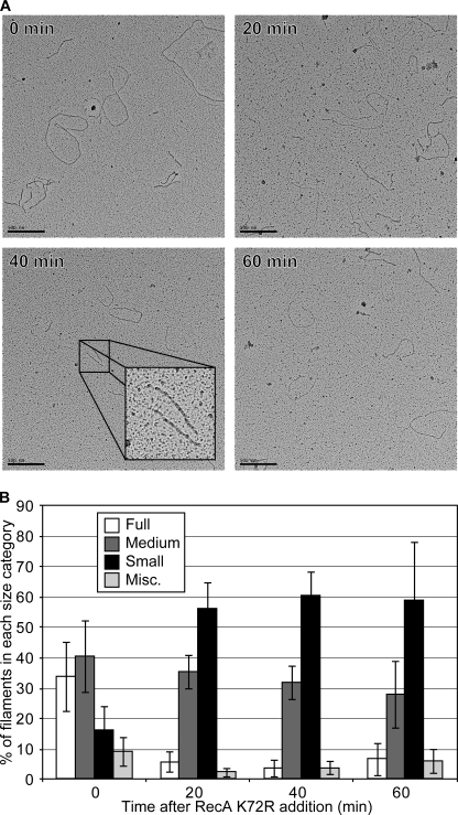
FIGURE 7.
RecA filaments decrease in length after wild type RecA bound to cssDNA is challenged with RecA K72R. Wild type RecA filaments formed on cssDNA were challenged with varying concentrations of RecA K72R as in Fig. 6, and the resultant filaments were imaged by EM as described under “Experimental Procedures.” The concentrations of DNA, RecA, ATP, and SSB are the same as in the legend to Fig. 6B. A, images of RecA filaments before, 20, 40, and 60 min after the addition of RecA K72R were obtained at 15,000× magnification. B, more than 400 filaments at 0, 20, 40, and 60 min after RecA K72R addition were visually inspected and grouped into the same length categories as Fig. 5B and as described under “Experimental Procedures.” Bars represent the average percentage of RecA filaments in each length category as counted from four randomly selected squares on carbon grids containing spread reaction samples from each time point. Error bars represent 1 S.D. from the mean.

