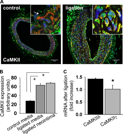
FIGURE 1.
CaMKII expression after carotid ligation. A, cross section of an untreated control carotid artery of a C57Bl/6 mouse (left panel) shows CaMKII immunoreactivity in the endothelium and media. 14 days of carotid ligation, strong CaMKII immunoreactivity (green) is seen in the neointima (smooth-muscle actin red, DAPI blue). Inset: detail of the carotid media (control) and neointima (ligated) at ×63 magnification. Representative example is ∼2.0 mm proximal to the ligation. Scale bar, 50 μm. B, densitometric quantitation of CaMKII expression in the media of the untreated control and neointima and media of a ligated C57Bl/6 mouse carotid artery 14 days after ligation. The measurements are given in arbitrary units adjusted for area and background. C, CaMKII isoform mRNA expression in carotid arteries of C57Bl/6 mouse 14 days after ligation by qrtPCR. Five carotids were pooled per measurement; data are representative of three independent experiments (*, p < 0.05).

