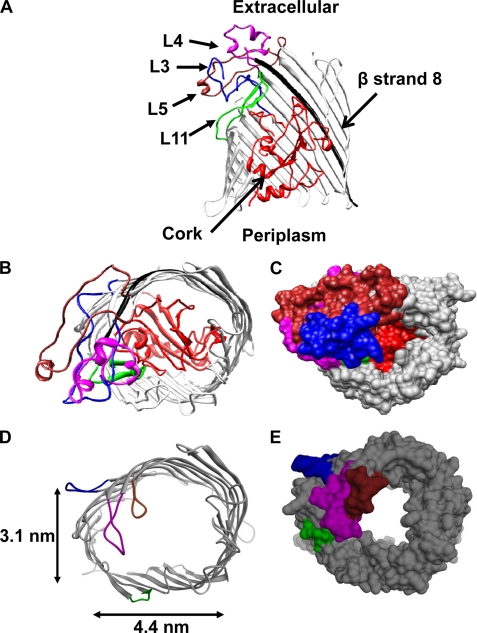
FIGURE 1.
Structure of the FhuA protein. A, ribbon diagram of the WT-FhuA protein (side view). Domains that were targeted for modifications in this study are the following: loops L3 (blue), L4 (magenta), L5 (brown), L11 (green), the first 160 amino acids, the cork (red), and strand β8 in the barrel (black). B, extracellular view of the WT-FhuA protein. C, surface representation of the extracellular view of the WT-FhuA protein, showing that the cork domain completely fills the pore lumen. D, ribbon diagram of the engineered FhuAΔC/Δ4L protein viewed from the extracellular side. E, surface representation of the engineered FhuAΔC/Δ4L protein.

