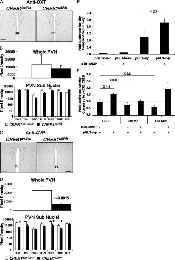
FIGURE 8.
CREB1ΔSIM1 mice have decreased in AVP expression. A, immunohistochemistry for OXT in CREB1lox/lox and CREB1ΔSIM1 mice. B, quantification of OXT immunohistochemistry in CREB1lox/lox and CREB1ΔSIM1 mice. C, immunohistochemistry for AVP in CREB1lox/lox and CREB1ΔSIM1 mice. D, quantification of AVP expression in CREB1lox/lox and CREB1ΔSIM1 brains. The data represent the pixel density in whole PVN and in the different subnuclei of the PVN and are expressed as the means ± S.E. *, p < 0.05. The mice were 33 weeks old, fed with chow diet, and housed at 22 °C (n = 4/group). PaAP, paraventricular hypothalamic nucleus, anterior parvicellular part; PaDC, paraventricular hypothalamic nucleus, dorsal cap; PaLM, paraventricular hypothalamic nucleus, lateral magnocellular part; PaMM, paraventricular hypothalamic nucleus, medial magnocellular part; PaMP, paraventricular hypothalamic nucleus, medial parvicellular part; PaPo, paraventricular hypothalamic nucleus, posterior part; PaV, paraventricular hypothalamic nucleus, ventral part; 3V, third ventricle. Bar scale, 100 μm. E, 293T cells were transfected with either pGL3 AVP-luc or pGL3-luc alone and a CMV-β galactosidase control. *, p < 0.05. F, 293T-cells were cotransfected with AVP-Luc, a CMV-β galactosidase control, and either pKCR2-CREB, pKCR2-CREMα, or pCDNA3.1-CREMτ2 in the presence or absence of 250 μm 8-bromo-cAMP (8-Br-cAMP).

