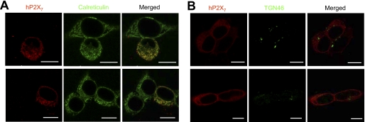
FIGURE 8.
Localization of intracellular hP2X7 proteins within the ER compartment. Shown are representative immunofluorescence confocal images of double staining for WT hP2X7 proteins (red) and calreticulin (green), an ER protein marker (A), and double staining for WT hP2X7 proteins (red) and TGN46 (green), a trans-Golgi network protein marker (B). Scale bars = 10 μm.

