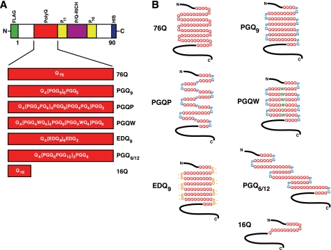
FIGURE 1.
Htt exon-1 polyQ and compact β polyQ proteins used for mammalian cell culture transfection experiments. A, domain structure of Htt exon-1 fragment, indicating polyQ region (red). Also shown are two Pro-repeat regions (yellow) and the P/Q-rich region (purple). An N-terminal FLAG tag (green) and a C-terminal His6 tag (blue) were engineered into each construct. The primary structure for each polyQ region is shown. B, predicted secondary structure of polyQ region in the expressed protein.

