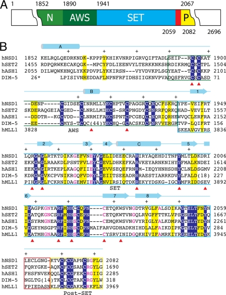
FIGURE 1.
Domain structure and sequence alignment of NSD1-CD. A, the domain structure of NSD1-CD. Each domain is represented by a colored box with the domain name indicated (N, N-terminal domain; P, post-SET domain). The numbers above and below the colored boxes indicate the numbers of the first and last residues of the domains, respectively. The red box indicates the post-SET loop connecting the SET and post-SET domains. B, structure-guided sequence alignment of the catalytic domains of human NSD1, SET2, ASH1, MLL1, and Neurospora Dim-5. Identical residues are indicated with white letters over a blue background, similar residues are highlighted in yellow, and those with all but one identical residue are shown in red. Different domains are enclosed in boxes colored as in A. A schematic diagram of the secondary structure elements of NSD1-CD is shown above the sequences, and every 10 residues is indicated with a + sign. Numbers to the left or right of the sequence indicate the numbering of the end residues. Note that the numbering for Dim-5 follows that of the PDB entry (1PEG). A red triangle below the sequences indicates the positions of missense mutations associated with Sotos syndrome.

