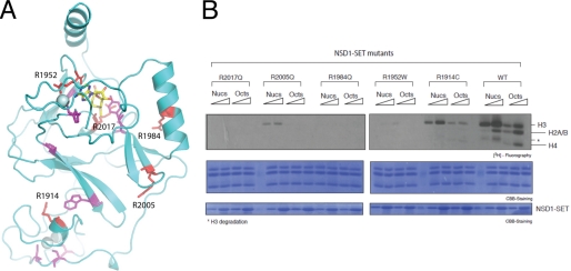
FIGURE 5.
Sotos syndrome missense mutations in NSD1-CD. A, amino acids at positions where the missense mutations of Sotos syndrome occur are shown as stick models (red and magenta), superimposed on a ribbon diagram of the NSD1-CD structure. AdoMet is shown as a stick model (yellow, carbon; blue, nitrogen; red, oxygen), and the spheres represent zinc ions. The residues shown in red correspond to arginines selected for in vitro mutagenesis and HKMTase assays. B, five arginine residues were individually changed to amino acids found in Sotos syndrome patients, and their HKMTase activities toward recombinant nucleosomes (Nucs) and histone octamers (Octs) were assayed using [3H]AdoMet as the methyl donor (top panel). The concentrations of NSD1-CD used in the assays were 0.04 and 0.2 μm, and those of nucleosomes and histone octamers were both 0.35 μm. Coomassie Brilliant Blue (CBB) staining of histones and the enzyme are shown in the middle and bottom panels, respectively.

