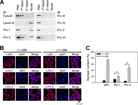
FIGURE 1.
PrxI and PrxII are located in the nucleus. A, immunoblot (IB) analysis of Prx isoforms in the subcellular fractions of HeLa cells. The α-tubulin and lamin B are cytosolic and nuclear markers, respectively. One representative blot of three experiments is shown. PNS, post-nuclear supernatant. B and C, immunostaining of PrxI and PrxII in HeLa cells treated either with or without leptomycin B (nuclear exportin-1 inhibitor). The NF-κB p65 protein is used as a positive control. Nuclei are labeled with DAPI (blue). One representative set of three experiments is shown. Data in the graph (C) are means ± S.D. of the percent of nuclear immunoreactive fluorescence versus total fluorescence obtained from 25 to 35 cells (n = 3; *, p < 0.005). LMB, leptomycin B.

