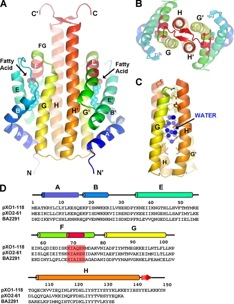
FIGURE 1.
Structure of the B. anthracis pXO1-118 sensor domain. A, ribbon representation of the dimer (side view), with termini and helices labeled (according to standard globin nomenclature). Helices are colored in spectral order (blue → orange) for each monomer. Labels for second monomer are primed. The KIAXER motif within helix F is colored red. Fatty acid is shown as cyan (methylene carbons) and red (carboxyl oxygens) spheres. B, same as in A, but orthogonal view looking down the 2-fold axis of the dimer. C, same view as in A, highlighting the dimer interface, with water molecules found only in a central region. D, sequence alignments of pXO1-118, pXO2-61, and the sensor domain of the B. anthracis sporulation kinase, BA2291. Secondary structure elements for pXO1-118 are indicated, as is the KIAXER motif.

