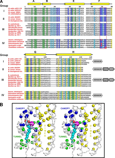
FIGURE 5.
Conservation of structure and function in the sensor domain family. A, structure-based sequence alignment and conservation of functional surfaces: fatty acid-binding pocket (hydrophobic tunnel), highlighted in green; canopy creating fatty acid/chloride-binding site (blue); dimer interface (yellow); and other conserved residues important for structural integrity (cyan). Sequences are grouped into four classes (I–IV) as defined under “Results.” Full names of the organisms and genes are as follows. Group I: B. anthracis pXO1-118 (NP_052814), B. anthracis pXO2-61 (NP_653040), B. cereus G2941 (ZP_00236329), B. cellulosilyticus DSM 2522 hypothetical protein BcellDRAFT_3961 (ZP_06365458). Group II: B. anthracis BA2291 (NP_844676.1), B. cereus sensor histidine kinase (YP_083662.1), B. thuringiensis sensor histidine kinase (YP_036402.1). Group III: B. coahuilensis m4-4 sensor histidine kinase (ZP_03225478), Bacillus sp. SG-1 histidine protein kinase (ZP_01860026), Brevibacillus brevis NBRC 100599 probable two-component sensor histidine kinase (YP_002774563), Geobacillus thermoglucosidasius C56-YS93 histidine kinase (ZP_06810042), Geobacillus sp. C56-T31 histidine kinase (YP_003671552), Bacillus sp. NRRL B-14911 sensor histidine kinase (ZP_01170643), Bacillus sp. SG-1 (ZP_01860665); Group IV: Anaeromyxobacter dehalogens 2CP-C conserved hypothetical protein (ACL66915.1), Chlorobium luteolum DSM 273 hypothetical protein Plut_0146 (ABB23036.1), Chloroherpeton thalassium ATCC 35110 conserved hypothetical protein (ACF14428.1), Syntrophus aciditrophicus SB hypothetical cytosolic protein (ABC79005.1), Candidatus Solibacter usitatus Ellin6076 hypothetical protein Acid_3380 (ABJ84353.1). B, stereo image of a pXO1-118 monomer, together with the dimer interface of the second monomer. Main chain (N-Cα-C) is shown as ribbon. The Cα positions of residues highlighted in A are shown as spheres, using the same color scheme. Fatty acid is in cyan and red, chloride ion in magenta.

