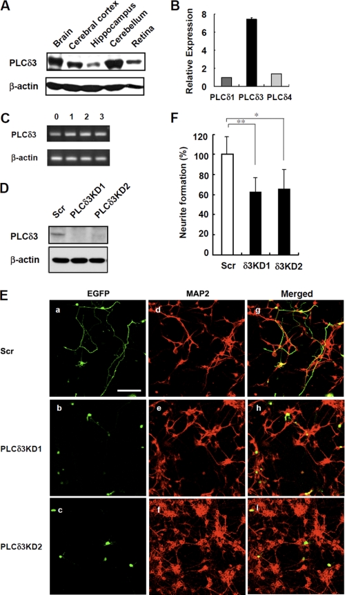
FIGURE 1.
PLCδ3 regulates neuronal outgrowth in cerebellar granule cells. A, expression of PLCδ3 was analyzed in the whole brain, cerebral cortex, hippocampus, cerebellum, and retina by Western blots with a monoclonal anti-PLCδ3 antibody that specifically recognizes the X-Y region of PLCδ3. β-Actin is used as the loading control. B, expression levels of PLCδ isozymes in isolated cerebellar granule cells were determined by qRT-PCR. The relative amounts are represented as the ratio to that of PLCδ1 mRNA. C, PLCδ3 was expressed throughout the 3-day culture of cerebellar granule cells isolated from P2–3 ICR mice. The amounts of PLCδ3 were examined by RT-PCR. D, endogenous PLCδ3 was effectively down-regulated by transfection with pGSU6-GFP vector carrying scrambled sequence or two distinct shRNA targeting mouse PLCδ3 sequence in Neuro2a cells. The expression levels of PLCδ3 were determined by Western blots. E, PLCδ3 knockdown caused significant inhibition of neurite outgrowth of cerebellar granule cells. 18 h after plating, cultured granule cells were transfected with control pGSU6-GFP (Scr, panels a, d, and g) or PLCδ3 shRNA expressing pGSU6-GFP (PLCδ3KD1, panels b, e, and h, or PLCδ3KD2, panels c, f, and i) and cultured for another 48 h. Note that PLCδ3 knockdown caused lower population of the granule cells with neurite compared with GFP-positive control cells (panels a–c). Middle (panels d–f) shows the granule cells stained with anti-MAP2 antibody, and right (panels g–i) shows the merged image. Scale bar, 100 μm. F, percentage of neurite formation in both GFP and MAP2-positive cells was evaluated and represented as a relative value. The rate of neurite outgrowth was determined by the population of neurons showing more than two cell body lengths. The data indicate the mean ± S.D. of five experiments obtained from average number of 600 GFP-positive cells counted for each transfection by microscopy. *, p < 0.05; **, p < 0.01 (Tukey's multiple comparison test).

