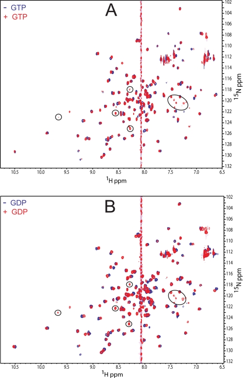
FIGURE 5.
NMR detection of guanine nucleotide interactions with Rv1739 STAS. A, two-dimensional 1H-15N HSQC spectra of Rv1739c STAS in the absence (blue contours) and presence of 20 mm GTP (red contours). B, two-dimensional 1H-15N HSQC spectra of Rv1739c STAS in the absence (blue contours) and presence of 20 mm GDP (red contours). Circled resonances indicate residues detected only in the presence of nucleotide. The guanine proton peak is at ∼8.07 ppm.

