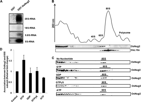
FIGURE 6.
Association between OsNug2 and ribosomal subunits in the presence of various nucleotides. A, rRNA binding assay. GST or GST-OsNug2 was mixed with total Oc cell lysate and the mixture incubated with glutathione-agarose 4B. Following centrifugation, the supernatant was discarded. Residual precipitates were treated with phenol to obtain rRNAs. Isolated rRNAs were separated on a 15% formaldehyde-agarose gel and then hybridization was performed using specific rRNAs as probes. B, whole cell lysates from Oc cells were sedimented through 7–47% sucrose gradients. The UV profiles at OD260 nm of individual fractions show 40S and 60S subunits, 80S whole ribosomes and polysomes. The upper column under the UV profile graph represents that after the TCA precipitation, an equal volume of proteins from each fraction were analyzed by SDS-PAGE and immunoblotted with anti-OsNug2 antibody. In the next column, the 60S ribosomal subunit was detected with anti-human L10a antibody, instead of OsL10a. C, ribosomal subunits in the presence of 2 mm GTP, GDP, GTPγS, or ATP, were sedimented through 7–47% sucrose gradients, as described in B. D, relative fold-changes of association between OsNug2 and 60S ribosomal subunits. The data represent the means of three independent measurements. Error bars represent mean ± S.D.

