
FIGURE 2.
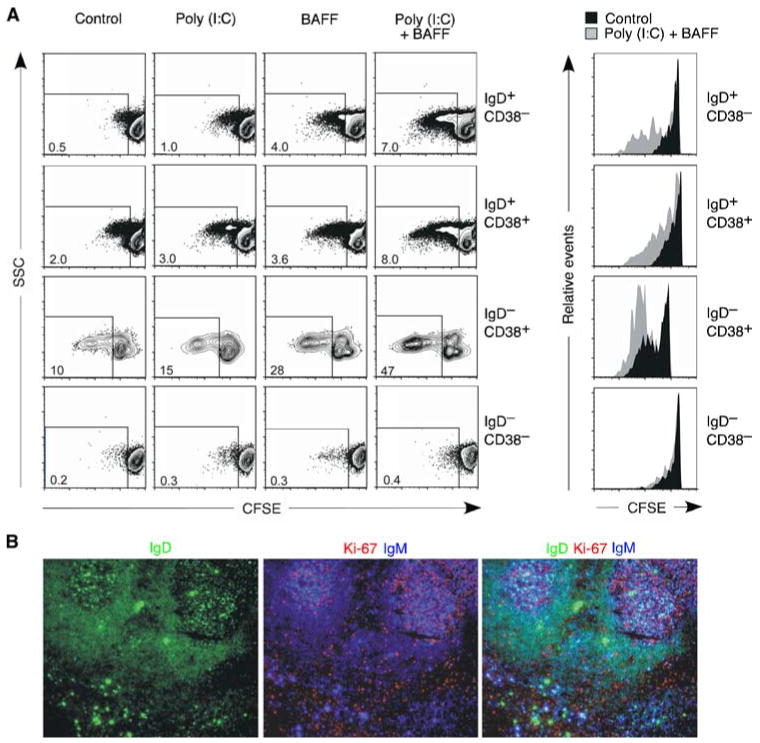
Upper respiratory mucosa B cells proliferate in response to dsRNA. A, Flow cytometric analysis of CFSE-labeled tonsillar naive IgD+CD38−, founder GC/activated IgD+CD38+, GC IgD−CD38+, and memory IgD−CD38− B cell subsets cultured with or without poly(I:C) and/or BAFF. Dot plots indicate percentage of B cells showing reduced CFSE expression over a 6-day culture. Histograms show CFSE dilution by control unstimulated (solid filled histograms) or poly(I:C)- and BAFF-stimulated (solid gray histograms) B cells gated as shown in the dot plots on the left. B, Immunofluorescence analysis of tonsillar mucosa stained for IgD (green), Ki-67 (red), and IgM (blue). Original magnification, ×5. Figure shows one of three experiments yielding similar results.