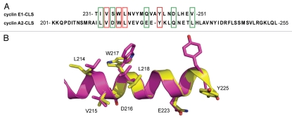
Figure 1.
The CLS domains of cyclins A and E are similar. (A) Amino acid sequence alignment for cyclin E1 and cyclin A2 CLSs. Identical residues are boxed in red and similar residues are boxed in green. (B) Ribbon diagram showing superimposition of cyclins E (yellow, PDB ID 1W98) and A (magenta, PDB ID 1VIN) CLSs in the conserved region. Identical and similar residues are represented as stick models.

