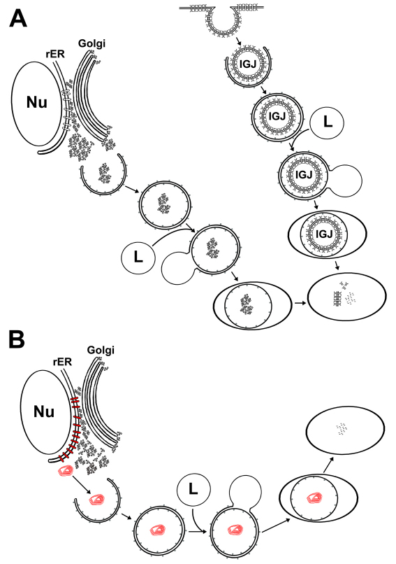
Fig. 11.
Diagram of the hypothesized autophagic process for connexins. Proposed sequence of events involved in the degradation of wild-type (A) or mutant (B) connexins through autophagy based on results presented in this study and published ultrastructural studies. (A) Wild-type connexins in the cytoplasm present in vesicles from the secretory pathway or in internalized gap junctions (IGJs) are entrapped by the LC3-containing isolation membrane into an autophagosome. The autophagosome then fuses with a lysosome (L) forming an autolysosome. Lysosomal enzymes degrade lipid and protein components of the entrapped material. Differences in cell types and the kinetics of the different steps might determine the frequency with which connexin-containing structures can be observed in autophagosomes. (B) Cytoplasmic accumulations of mutant connexin (red) are entrapped by the isolation membrane into an autophagosome that then fuses with a lysosome (L) forming an autolysosome. After starvation, the mutant connexin in accumulations is degraded by lysosomal proteases. Under normal growth conditions, the levels of constitutive autophagy are insufficient to degrade the mutant protein and might contribute to the formation of large accumulations by bringing small accumulations together. LC3 is represented by gray dots on membrane surfaces. Nu, nucleus; rER, rough endoplasmic reticulum.

