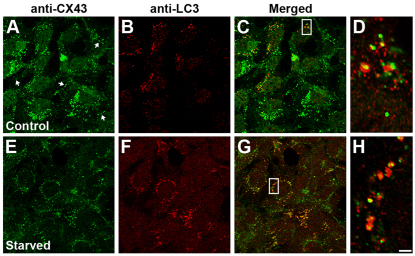
Fig. 6.
LC3 colocalizes with CX43 in mouse embryonic fibroblasts. (A–H) Confocal images show the immunolocalization of CX43 (A,E) and LC3 (B,F) in mouse embryonic fibroblasts incubated under control conditions (A–D) or starved for 1 hour (E–H). Colocalization of the two signals is shown in the merged panels (C and G). The boxed regions are shown at higher magnification after deconvolution in D and H. Scale bar: 16 μm (A–C,E–G) and 2 μm (D,H).

